

An ear flush under anesthesia cleans the whole ear canal with a video-otoscope, so the eardrum can be checked and ear medicine reaches the skin of the canal. At Saari Animal Clinic in Vaasa it costs €449–489 depending on your pet’s weight (under or over 15 kg). Anesthesia and pain relief are included; microscopy of the ear samples is charged separately.
Ear infections are among the most common reasons dogs see a vet. A hand-held otoscope is enough for most ear examinations, but swelling and discharge can hide the eardrum. In video-otoscopy, a thin endoscope with a camera shows a magnified view of the ear canal on a monitor.
Why Video-Otoscopy?
Thanks to far better magnification and lighting than a hand-held otoscope provides, even small eardrum tears can often be seen once the canal is clean. Fine instruments pass through the scope, so examination, sampling and treatment happen under a single anesthetic.
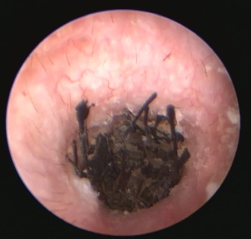
When Does a Dog or Cat Need an Ear Flush?
Most ear infections are diagnosed at an ordinary visit. Video-otoscopy is recommended when an infection keeps returning or resists treatment; the canal is blocked with discharge or the eardrum cannot be seen; or a foreign body (such as a grass awn), a growth, a ruptured eardrum or a middle-ear infection is suspected. In cats, it is also used to remove inflammatory polyps, benign middle-ear growths that can reach the ear canal.
How Is the Ear Flush Done?
Badly swollen canals may first need 1–3 weeks of anti-inflammatory medicine. During general anesthesia with pain relief, a breathing tube protects your pet’s airway, because flushing fluid can drain through an open eardrum into the throat. The canal is flushed clean. Samples are examined under the microscope in our own laboratory and, when needed, sent for bacterial culture. Foreign bodies and polyps can be removed and growths biopsied. If the eardrum is intact but middle-ear disease is suspected, the vet may make a small opening in it (myringotomy) to sample and flush the middle ear. The opening usually heals within about five weeks if it is kept free of infection.
Aftercare and Possible Side Effects
We give pain relief as needed and often leave a long-acting medicated gel in the cleaned canal. The procedure, especially opening the eardrum or removing a polyp, can cause head tilt and loss of balance, a drooping eyelid and a small pupil (Horner’s syndrome, more common in cats), facial paralysis or hearing loss. These signs usually pass within a few weeks but can be permanent. If you notice them, stop any ear drops and contact us the same day. In one study, polyps removed through the scope regrew in about one in seven cats.
Chronic Ear Infections and Their Challenges
In dogs, recurring ear infections almost always have an underlying cause, most often allergy. Repeated inflammation narrows the canal, microbes shelter in a slimy biofilm, hard-to-treat bacteria such as Pseudomonas become more common, and the middle ear is often infected too. A flush clears the biofilm so medication reaches the skin of the canal, but the underlying cause must still be found and controlled.
How Much Does an Ear Flush Cost?
At Saari Animal Clinic, an ear flush under general anesthesia with video-otoscopy costs €449–489, depending on your pet’s weight (under or over 15 kg). The price includes the anesthesia and pain relief. Microscopy of the ear samples, outside laboratory tests (bacterial culture, biopsies) and take-home medicines are charged separately. An ordinary ear examination with a sample checked under the microscope costs €137.79, including the visit fee. See our price list.
When Should You Contact Us?
Book an appointment if your pet keeps scratching its ears or shaking its head, its ears smell or have discharge, or infections keep returning. Contact us the same day if your pet tilts its head or walks unsteadily, its eyes flicker from side to side, one side of its face droops, or it is clearly in pain. Call (06) 321 7300 or use our online booking. Read more: Ear Infections in Dogs.
Sources
- Cole L.K. (2004): Otoscopic evaluation of the ear canal. Veterinary Clinics of North America: Small Animal Practice 34(2):397–410. pubmed.ncbi.nlm.nih.gov/15062615
- Cole L., Nuttall T. (2021): When and how to do a myringotomy – a practical guide. Veterinary Dermatology 32(3):302–e82. pubmed.ncbi.nlm.nih.gov/33955092
- Nuttall T. (2023): Managing recurrent otitis externa in dogs: what have we learned and what can we do better? Journal of the American Veterinary Medical Association 261(S1):S10–S22. pubmed.ncbi.nlm.nih.gov/37019436
- Greci V. ym. (2014): Per-endoscopic trans-tympanic traction for the management of feline aural inflammatory polyps: a case review of 37 cats. Journal of Feline Medicine and Surgery 16(8):645–650. pmc.ncbi.nlm.nih.gov/PMC11164151
- Secker B. ym. (2023): Pseudomonas spp. in Canine Otitis Externa. Microorganisms 11(11):2650. pmc.ncbi.nlm.nih.gov/PMC10673570
- Hoff S. (2025): Otitis Externa in Animals. Merck Veterinary Manual. merckvetmanual.com/otitis-externa-in-animals
- Hoff S. (2025): Otitis Media and Interna in Animals. Merck Veterinary Manual. merckvetmanual.com/otitis-media-and-interna-in-animals