

Tooth resorption is a disease in which the body’s own cells (odontoclasts) gradually destroy the hard tissues of a tooth — cementum, dentin, and enamel. It usually starts on the root, below the gum line, and gets worse over time. It is most common in cats but is increasingly recognized in dogs. Once a lesion opens into the mouth, it is often painful — yet most affected cats show no obvious signs, so the disease easily goes unnoticed.
How Common Is Tooth Resorption?
Depending on the population and how the cats were examined, tooth resorption has been found in 28.5–67% of cats, and it becomes more common with age. In a University of Helsinki owner survey of 8,115 Finnish cats, a vet had diagnosed tooth resorption in 21% of the 944 cats examined or treated under sedation for oral or dental disease. Cornish Rex, European (European Shorthair), and Ragdoll cats had a higher risk, and Exotic and Persian cats a lower one, though breed findings vary between studies. In dogs, full-mouth radiographs showed tooth resorption in 54% of 224 dogs at a US university dental service; in a Korean study of small-breed dogs, it became more common from about 9 years of age. In cats, the lower premolars are affected most often; in dogs, premolars and molars are commonly affected.
Types — Why Radiographs Determine Treatment
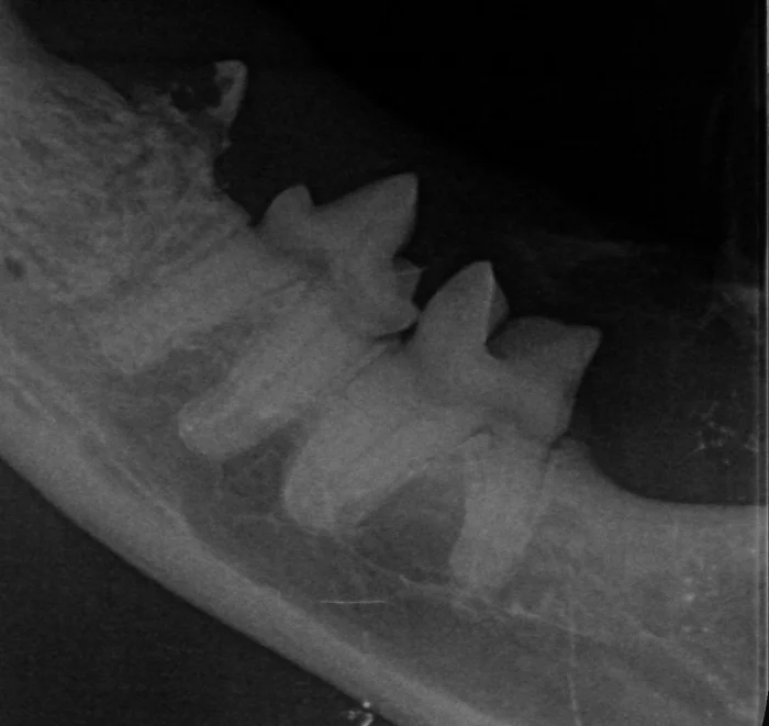
In cats, tooth resorption is divided into three types, and telling them apart requires dental radiographs. In Type 1, parts of the tooth are destroyed but not replaced: the radiograph shows dark defects, while the root outline and the periodontal ligament — the thin attachment between root and jawbone — are still visible. Type 1 is linked to inflammation, especially periodontitis (gum disease). In Type 2, the root is gradually replaced by bone-like tissue and fuses with the jawbone (ankylosis): the periodontal ligament narrows or disappears, and the root fades into the bone like a “ghost.” The cause of Type 2 is unknown. In Type 3, both patterns occur in the same tooth — for example, one root of Type 1 and another of Type 2.
Symptoms — Hidden Pain
Tooth resorption is often a “silent” disease: a lack of signs at home does not mean the teeth are pain-free. Signs you may notice include dropping food, tilting the head or chewing on one side only, swallowing food without chewing, preferring soft food, drooling, jaw chattering, pawing at or rubbing the mouth, head shaking, grooming less, hiding, or playing less. Many pets keep eating despite the pain. On examination, the vet may see red or pink gum tissue growing into a defect at the gum line, inflamed gum around a single tooth, or missing teeth.
Diagnosis — Why Dental Radiographs Are Needed
Because lesions usually start below the gum line, one that is visible in the mouth is often already advanced. A reliable diagnosis needs a thorough examination under general anesthesia — every tooth checked with a fine dental probe — together with dental radiographs. In a Danish study of 144 unowned cats, the probe alone found just over a third of affected teeth; radiographs found about four in five. Radiographs also show the type, which determines the treatment. At Eläinklinikka Saari, we radiograph the whole mouth at every dental treatment under anesthesia — not only the problem teeth. A “missing” tooth is not always gone: its resorbing remains may be hidden beneath the gum.
Treatment — Type Determines the Method
Once started, a lesion keeps progressing and no medicine has been shown to stop it, so treatment aims to remove the source of pain. In Type 1, the whole tooth — crown and every root — is extracted surgically. For advanced Type 2 lesions, guidelines accept crown amputation: the crown is removed and the gum stitched closed, leaving in place root remnants that are turning into bone and are often very difficult or impossible to extract. This is suitable only if the radiograph shows no remaining periodontal ligament and there are no signs of periodontitis, infection inside the tooth (endodontic disease), or stomatitis (widespread mouth inflammation); otherwise the tooth is extracted. In Type 3, the Type 1 root is extracted, while a Type 2 root may be left in place, as in crown amputation. An early, painless lesion confined to the root can sometimes be monitored with repeat radiographs instead. Fillings do not stop the disease: in one study, only a third of 154 filled cat teeth showed no further loss of tooth structure after six months or more. At Eläinklinikka Saari, we perform extractions and crown amputations, and one of our vets has additional training in veterinary dentistry.
After Treatment
The mouth can be sore for the first days after extractions, so your pet goes home with pain relief medication. If the gum was stitched, we check it after about five days. Once a cat has had tooth resorption, new lesions are likely in other teeth, so feline dental guidelines advise repeat dental radiographs, ideally yearly. These need general anesthesia, so your vet will discuss timing with you.
Can Tooth Resorption Be Prevented?
There is no proven way to prevent tooth resorption, because the cause of most lesions is unknown — excess dietary vitamin D has been suggested as a cause but not confirmed. Type 1 lesions, however, are linked to gum disease: treating periodontitis is advised to help prevent new ones, and good home dental care helps keep the gums healthy. We recommend a mouth check at least once a year at a routine visit, for example with vaccinations; it cannot show root lesions, but it can reveal signs that call for radiographs under anesthesia. Book an appointment by calling (06) 321 7300.
Sources
- Niemiec B. ym. (2026): World Small Animal Veterinary Association global dental guidelines. Journal of Small Animal Practice, doi:10.1111/jsap.70057. pubmed.ncbi.nlm.nih.gov
- Lobprise H. ym. (2025): 2025 FelineVMA feline oral health and dental care guidelines. Journal of Feline Medicine and Surgery 27(11). pmc.ncbi.nlm.nih.gov
- Gorrel C. (2015): Tooth resorption in cats: pathophysiology and treatment options. Journal of Feline Medicine and Surgery 17(1):37–43. pmc.ncbi.nlm.nih.gov
- Vapalahti K. ym. (2024): A large case-control study indicates a breed-specific predisposition to feline tooth resorption. The Veterinary Journal 305:106133. pubmed.ncbi.nlm.nih.gov
- Peralta S. ym. (2010): Radiographic evaluation of the types of tooth resorption in dogs. American Journal of Veterinary Research 71(7):784–793. pubmed.ncbi.nlm.nih.gov
- Bae J. ym. (2025): Prevalence and classification of tooth resorption in small-breed dogs: a retrospective study using full-mouth intraoral radiographs. The Veterinary Journal 313:106377. pubmed.ncbi.nlm.nih.gov
- Eriksson J. ym. (2024): Accuracy of three diagnostic tests to detect tooth resorption in unowned unsocialised cats in Denmark. Journal of Small Animal Practice 65(6):387–393. pubmed.ncbi.nlm.nih.gov
- Lyon K.F. (1992): Subgingival odontoclastic resorptive lesions. Classification, treatment, and results in 58 cats. Veterinary Clinics of North America: Small Animal Practice 22(6):1417–1432. pubmed.ncbi.nlm.nih.gov
- Mulherin B.L. (2024): Tooth Resorption in Small Animals. MSD Veterinary Manual. msdvetmanual.com
- American Veterinary Dental College (AVDC): Nomenclature — tooth resorption stages and types. avdc.org