

An unerupted tooth is a tooth that has never broken through the gum into the mouth — it stays hidden under the gum or inside the jawbone. Unerupted teeth are seen particularly often in brachycephalic (short-nosed) breeds. A fluid-filled dentigerous cyst can develop around the crown of an unerupted tooth and gradually enlarge, destroying the surrounding jawbone. Because the tooth cannot be seen, the problem often goes unnoticed until the damage is significant — unless dental X-rays are taken. At our clinic, we take dental X-rays and surgically remove unerupted teeth and dentigerous cysts.
What Is a Dentigerous Cyst?
While a tooth develops, its crown is covered by a thin layer of tissue called the reduced enamel epithelium. Normally this layer merges with the gum as the tooth erupts. If the tooth stays buried, fluid can collect between this layer and the crown, forming a cyst. As fluid builds up, the cyst enlarges and the surrounding bone is gradually broken down (resorbed). Studies report that roughly 30–45% of unerupted teeth in dogs have a cyst around them. The tooth most often affected is the lower first premolar — the first small tooth behind the lower canine.
Symptoms — Often None at First
A dentigerous cyst usually causes no visible signs in the early stages — this is what makes it so insidious. The main clue is a “missing” tooth: a tooth that should be there but cannot be seen. Some dogs develop a soft swelling of the gum or jaw. A growing cyst can damage the roots of neighboring teeth, which may then die and become discolored. In severe cases, the cyst weakens the jaw so much that the jaw breaks during normal use (a pathological fracture). Even a dog with a normal number of visible teeth can have a hidden extra (supernumerary) tooth, which can also form a cyst.
At-Risk Breeds
Unerupted teeth and dentigerous cysts are clearly overrepresented in short-nosed breeds; in one study, brachycephalic dogs with unerupted teeth had about three times the odds of a cyst compared with non-brachycephalic dogs. The exact cause is unclear: an Italian study found that crowding in the jaw could explain only a few cases, and it is not yet known whether there is an inherited breed tendency. In an American study, Boxers, Pugs, Shih Tzus, and Boston Terriers were the breeds most likely to have these cysts. In a Finnish study of 192 dogs with these cysts, Tibetan Spaniels (30%) and Boxers (25%) made up more than half of the cases, followed by Chihuahuas and Pugs. Any breed can be affected, however. In cats, unerupted teeth and dentigerous cysts are rare but do occur — the same principles apply.
Diagnosis — Dental X-Rays Are Essential
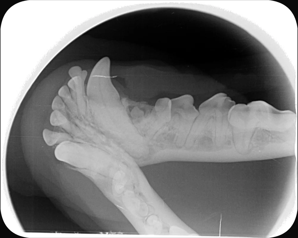
An unerupted tooth and its cyst cannot be seen by looking in the mouth — they are found with dental X-rays (or occasionally CT). On the X-ray, a cyst appears as a well-defined, round or oval dark (radiolucent) area around the crown of the buried tooth. The X-ray also shows how much bone has been lost and whether neighboring teeth are affected. The WSAVA dental guidelines recommend X-raying every “missing” tooth. The adult teeth come in at about 4–7 months of age, so a tooth can only be called missing once they are in. At puppy check-ups we count the teeth, and once the adult teeth have come in, we X-ray any tooth that is missing. We also take full-mouth X-rays at every dental procedure under general anesthesia: in dogs referred for dental treatment, X-rays of teeth that looked normal revealed clinically important findings in about 28% of dogs (Verstraete et al. 1998).
Treatment — Surgical Removal
Treatment is surgical removal of the unerupted tooth together with the entire cyst lining, which is carefully scraped out (curettage) so that none of it is left behind. A flap of gum and underlying tissue (a mucoperiosteal flap) is raised, the bone over the tooth is opened if needed, the tooth and cyst lining are removed, and the gum is sutured closed. Neighboring teeth badly damaged by the cyst may also need to be removed. According to the 2020 WSAVA dental guidelines, larger defects may benefit from a bone graft, and new bone typically fills the cavity within about three months. We recommend a follow-up dental X-ray to confirm that the bone has healed. A cyst should be removed once it is found, because untreated cysts tend to keep growing. Even when there is no cyst yet, we usually recommend removing an unerupted tooth to prevent one; in some dogs, regular X-ray monitoring can be an alternative. At Eläinklinikka Saari, we perform these surgeries ourselves — book a dental examination and we will assess the situation.
Prognosis
When a dentigerous cyst is found early and its lining is removed completely, the prognosis is excellent: in an Italian study, surgery was successful in every case that was followed up and no cyst recurred, although many of the follow-up periods were short. The outlook is more guarded if the cyst has grown large or destroyed a lot of bone, or if the jaw has already fractured. No case of a dentigerous cyst turning into cancer has been confirmed in dogs: in a large Finnish study, none of the 208 cysts examined showed malignant change. Tumors next to unerupted teeth have, however, occasionally been reported. Early diagnosis matters most: dental X-rays find unerupted teeth while any cyst is still small, or before one has formed.
Sources
- Babbitt S.G., Krakowski Volker M., Luskin I.R. (2016): Incidence of radiographic cystic lesions associated with unerupted teeth in dogs. Journal of Veterinary Dentistry 33(4):226–233. pubmed.ncbi.nlm.nih.gov/28218030
- Heinze C.S., Miles J.E., Gawor J., Kortegaard H.E. (2026): A cross-sectional radiographic study on the prevalence and distribution of dentigerous cysts in unerupted teeth in adult dogs. Journal of Small Animal Practice 67(5):422–431. pubmed.ncbi.nlm.nih.gov/41888382
- Bellei E., Ferro S., Zini E., Gracis M. (2019): A clinical, radiographic and histological study of unerupted teeth in dogs and cats: 73 cases (2001–2018). Frontiers in Veterinary Science 6:357. pmc.ncbi.nlm.nih.gov/PMC6856145
- Hutt J.P., Volker M.K., Sánchez M.D. (2022): Correlation between radiographic and histopathologic findings associated with unerupted teeth in dogs. Journal of Veterinary Dentistry 39(4):330–336. pubmed.ncbi.nlm.nih.gov/35711165
- Vesterinen S., Lönnberg L., Junnila J.J.T., Luotonen N., Kuntsi H. (2024): Clinical and histopathological study of 279 dentigerous cysts in 192 dogs (2012–2022). Frontiers in Veterinary Science 11:1412089. pmc.ncbi.nlm.nih.gov/PMC11156172
- Verstraete F.J.M., Kass P.H., Terpak C.H. (1998): Diagnostic value of full-mouth radiography in dogs. American Journal of Veterinary Research 59(6):686–691. pubmed.ncbi.nlm.nih.gov/9622735
- Niemiec B. et al. (2020): World Small Animal Veterinary Association Global Dental Guidelines. Journal of Small Animal Practice 61(7):E36–E161. pubmed.ncbi.nlm.nih.gov/32715504
- Merck Veterinary Manual, Dog Owners: Dental Development of Dogs. merckvetmanual.com