

Cranial cruciate ligament (CCL) disease is one of the most common causes of hind-leg lameness in dogs. The ligament usually weakens gradually and often tears partially before rupturing completely. The unstable knee is painful, and osteoarthritis develops in the joint. TTA (tibial tuberosity advancement) stabilizes the knee surgically without replacing the ligament.
How Does TTA Work?
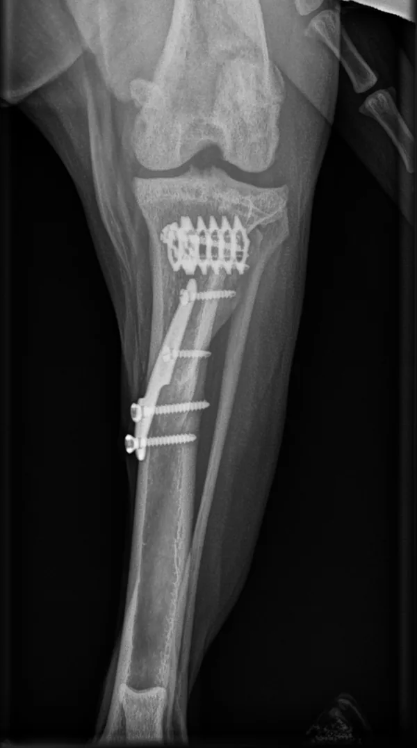
During TTA, the tibial tuberosity — the bony point below the knee where the kneecap tendon attaches — is cut and moved forward by an amount planned from X-rays. It is then fixed in its new position with a titanium cage and a plate secured with a fork and screws. As a result, the kneecap tendon runs at about 90 degrees to the joint surface at the top of the shin bone. This counteracts the force that pushes the shin bone forward when the dog bears weight (cranial tibial thrust). We also check the menisci (the knee’s cartilage cushions) and remove or treat a torn meniscus. At our clinic, TTA is performed by veterinarian Pamela Kvarngård, who has extensive experience in orthopedic procedures.
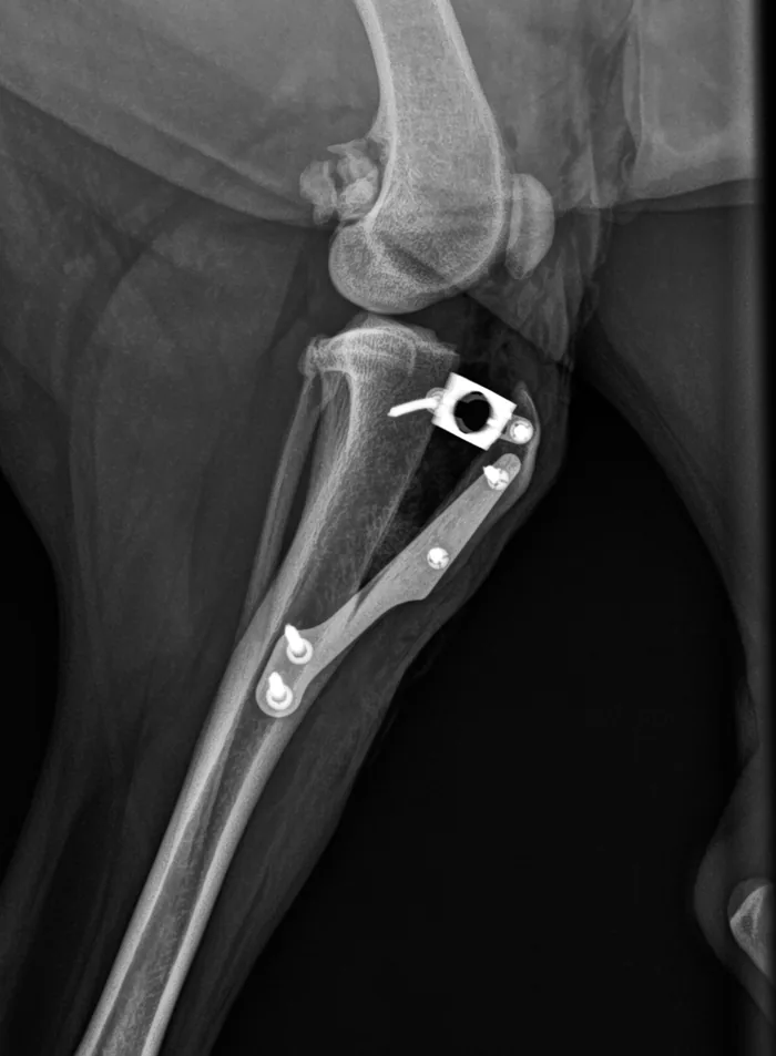
TTA or Lateral Suture?
We offer two surgical treatments for cruciate ligament disease: TTA and the lateral suture. The lateral suture stabilizes the knee with strong synthetic material placed outside the joint to mimic the ligament, whereas TTA permanently changes the forces acting on the knee. The lateral suture suits cats and small dogs, and often medium-sized dogs and older, calmer large dogs as well. TTA suits active dogs and large dogs. We always assess your dog individually to decide which method suits it best.
Recovery
Your dog goes home with pain medication and a written home-care plan: leash walks only, with no running, jumping or free play until the bone has healed. Stitches come out at 12–14 days; a follow-up X-ray at 6–8 weeks checks bone healing before exercise is increased gradually. By 3–4 months most dogs walk with no limp or only a mild one, and improvement can continue for up to a year. We always recommend animal physiotherapy, which can speed recovery. An animal physiotherapist also sees patients at our clinic. In studies, about 9 in 10 owners rate the result as good or excellent.
Risks and Complications
Most studies report complications in about 1 in 10 to 1 in 3 dogs. Roughly 1 in 10 dogs has a more serious complication, often needing another operation, but most such problems can be treated successfully. Complications include infection, fracture of the tibial tuberosity or shin bone, implant failure, kneecap dislocation and a later meniscal tear (usually reported in 2–10% of knees). Osteoarthritis often still progresses over time, even after successful surgery. During surgery, we monitor anesthesia closely and give a multi-drug pain-relief infusion.
When Should You Contact Us?
If your dog limps on a hind leg, has trouble rising or sitting, or is stiff after rest, cruciate ligament disease is a common cause. Partial tears usually progress, and delaying treatment raises the risk of meniscal damage, so do not wait. Roughly a third to a half of dogs with the disease in one knee later develop it in the other knee — older dogs less often. Keep your dog lean, as excess weight is a risk factor. Book an orthopedic examination and we will recommend the most suitable method.
Sources
- American College of Veterinary Surgeons (ACVS). Cranial Cruciate Ligament Disease (animal-owner health topic). acvs.org
- Apelt D, Kowaleski MP, Boudrieau RJ (2007). Effect of tibial tuberosity advancement on cranial tibial subluxation in canine cranial cruciate-deficient stifle joints: an in vitro experimental study. Vet Surg 36(2):170–177. doi.org/j.1532-950X.2007.00250.x
- Edwards GA, Hosgood G, Hancock RB, Stubbs WP, Jackson AH (2016). Major complications associated with fork-based and screw-based tibial tuberosity advancement implants: 438 cases. Can Vet J 57(4):415–420. pmc.ncbi.nlm.nih.gov/PMC4790234
- Lafaver S, Miller NA, Stubbs WP, Taylor RA, Boudrieau RJ (2007). Tibial tuberosity advancement for stabilization of the canine cranial cruciate ligament-deficient stifle joint: surgical technique, early results, and complications in 101 dogs. Vet Surg 36(6):573–586. doi.org/j.1532-950X.2007.00307.x
- Steinberg EJ, Prata RG, Palazzini K, Brown DC (2011). Tibial tuberosity advancement for treatment of CrCL injury: complications and owner satisfaction. J Am Anim Hosp Assoc 47(4):250–257. doi.org/JAAHA-MS-5574
- Hoffmann DE, Miller JM, Ober CP, Lanz OI, Martin RA, Shires PK (2006). Tibial tuberosity advancement in 65 canine stifles. Vet Comp Orthop Traumatol 19(4):219–227. doi.org/s-0038-1633004
- Stein S, Schmoekel H (2008). Short-term and eight to 12 months results of a tibial tuberosity advancement as treatment of canine cranial cruciate ligament damage. J Small Anim Pract 49(8):398–404. doi.org/j.1748-5827.2008.00592.x
- MacDonald TL, Allen DA, Monteith GJ (2013). Clinical assessment following tibial tuberosity advancement in 28 stifles at 6 months and 1 year after surgery. Can Vet J 54(3):249–254. pmc.ncbi.nlm.nih.gov/PMC3573630
- Costa M, Craig D, Cambridge T, Sebestyen P, Su Y, Fahie MA (2017). Major complications of tibial tuberosity advancement in 1613 dogs. Vet Surg 46(4):494–500. doi.org/vsu.12649
- Hayes GM, Langley-Hobbs SJ, Jeffery ND (2010). Risk factors for medial meniscal injury in association with cranial cruciate ligament rupture. J Small Anim Pract 51(12):630–634. doi.org/j.1748-5827.2010.01003.x
- Grierson J, Asher L, Grainger K (2011). An investigation into risk factors for bilateral canine cruciate ligament rupture. Vet Comp Orthop Traumatol 24(3):192–196. doi.org/VCOT-10-03-0030
- Murphy CL, Niles J, Radasch RM (2024). The prevalence and risk factors of contralateral cranial cruciate ligament rupture in medium-to-large (≥15 kg) breed dogs 8 years of age or older. Vet Comp Orthop Traumatol 37(1):8–12. doi.org/s-0043-1771350